Short answer: a CBCT scan is a three-dimensional X-ray of your jaw. It lets bone height, width and density be measured, and it shows exactly where your nerve canal and sinuses sit. A standard dental X-ray cannot do any of that — which is why an implant plan built without 3D imaging is, in large part, an educated guess.
If you have been told you need a scan before an implant and quietly wondered whether it is a necessary step or an upsell, this is a fair question and it deserves a real answer.
A flat X-ray is a shadow. A 3D scan is a model.
A conventional dental X-ray — the bitewing, the periapical, the panoramic — projects your anatomy onto a flat image. Everything in the path of the beam is layered on top of everything else. It is genuinely useful for spotting decay, checking root shape, and seeing gross bone levels.
What it cannot show is depth.
CBCT stands for cone beam computed tomography. The machine rotates around your head and captures a volume rather than a picture. That volume can be sliced and viewed from any direction, and — this is the part that matters — it can be measured.
| Standard dental X-ray | CBCT scan | |
|---|---|---|
| What it produces | A flat, two-dimensional image | A three-dimensional volume you can slice and rotate |
| Bone height | Roughly visible | Measurable |
| Bone width (front-to-back) | Not visible at all | Measurable |
| Bone density | Not assessable | Assessable |
| Nerve canal position | Approximate | Located in three dimensions |
| Sinus floor position | Approximate | Located in three dimensions |
| Good for | Decay, root anatomy, general screening | Surgical planning inside bone |
Both have a place. A CBCT is not a better version of a bitewing; it answers a different set of questions.
The four things the scan is actually looking for
1. Is there enough bone, and where?
An implant needs bone around it on every side. A ridge can look perfectly adequate on a panoramic X-ray and turn out to be a narrow blade of bone once you see it in cross-section. Width is invisible in two dimensions, and width is frequently the limiting factor.
2. How dense is that bone?
Bone quality varies across the mouth, and it affects how firmly an implant sits when it is placed and how the healing period is planned. Denser bone in the lower jaw behaves differently from softer bone in the upper back jaw.
3. Where is the nerve?
In the lower jaw, the inferior alveolar nerve runs through a canal in the bone and supplies sensation to the lip and chin. Knowing precisely where it runs — and how much bone sits above it — is how the implant length is chosen. This is one of the clearest patient-facing reasons 3D imaging exists.
4. Where is the sinus?
In the upper back jaw, the maxillary sinus sits directly above the roots of the molars, and the floor of that sinus drops as bone is lost. The scan measures the remaining height, which is how it becomes clear whether a sinus lift or bone graft is part of the plan or not.
How the scan turns into a surgical plan
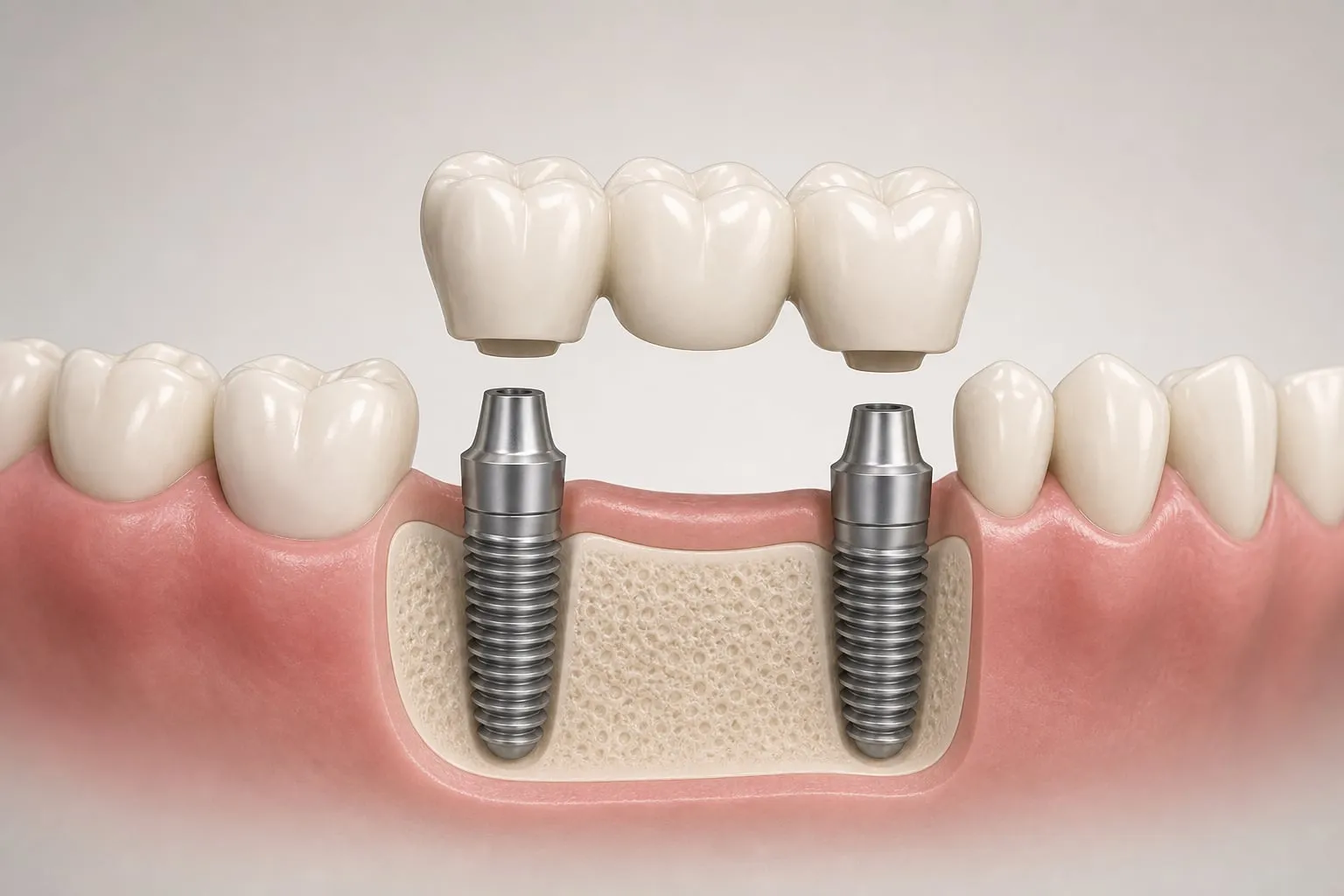
Once the volume exists, the implant can be positioned virtually before anything happens in your mouth. The planned size, depth and angle can be checked against the nerve, the sinus, the roots of neighboring teeth, and the shape of the bone.
Combined with digital scanning of your teeth, that plan can also account for where the finished tooth needs to sit — because an implant is not placed where there is convenient bone, it is placed where the restoration needs it, and the two have to be reconciled. You can read more about 3D imaging and implant planning, or see the technology we use.
The honest framing of the benefit: published comparisons generally report that guided placement achieves closer agreement with the planned angle and position than freehand placement does. The practical value of that accuracy is largest near critical anatomy — the nerve canal, the sinus floor, an adjacent root — and in anatomically complicated cases. It is a risk-reduction tool, not a claim that the implant itself is better.
Why a quote without imaging is a guess
This is the part with real money attached.
An implant estimate has to account for whether grafting is needed, how many implants are appropriate, what length and width they will be, and whether the site can take them at all. Every one of those depends on measurements that only exist in three dimensions.
A number produced before the scan is a placeholder. It can move considerably in either direction once the bone is actually visible — which is why a figure quoted over the phone, by anyone, should be treated as a starting point rather than a price.
For reference, single implants at our practice start at $1,995, which covers the consultation, the surgery, and the crown. Pricing may vary based on individual needs — contact the office for a personalized estimate. More detail on what drives the number is on our implant cost page and in what’s included in the $1,995 implant.
The radiation question
It is a reasonable thing to ask. Published effective-dose figures for dental cone beam scans sit well below those of a medical CT of the head, and are commonly described as comparable to a small number of days of ordinary background radiation, with the dose varying according to how large an area is scanned.
The more useful principle than any single number: imaging should be taken because it will change or confirm what is done, not by default. A scan that determines implant length, angle and whether you need grafting is doing real work.
The bottom line
A CBCT scan is how an implant plan stops being an estimate and becomes a set of measurements. It shows bone width and density, locates the nerve and the sinus, and lets the implant be positioned virtually before surgery. If you have been told you may not have enough bone, it is also how you find out whether that is actually true — see dental implants with bone loss and what determines candidacy.
Your first consultation visit is where the imaging and the conversation happen together.
Get the measurements before you make a decision
A free consultation gives you an exam, imaging, and a plan based on what is actually there. Request your free consultation or call (949) 773-4170.
This article is general education, not a diagnosis or medical advice. Individual results vary, and only a personalized evaluation can determine what imaging and treatment may be appropriate for you.
References: Radiation doses in dental radiology (IAEA) · Associated risks with periodontal x-rays or CBCT scans (Clinical Advances in Periodontics)